Structure of the eye
To understand how common conditions can affect the eyes and eyesight, it’s important to understand the eye’s structure and how it works. Sight is actually a series of events triggered by light. When light is reflected from an object, it passes through the pupil at the front of the eye and is focused by the lens onto the retina at the back. Cells in the retina convert this information into a signal that travels via the optic nerve to the brain.
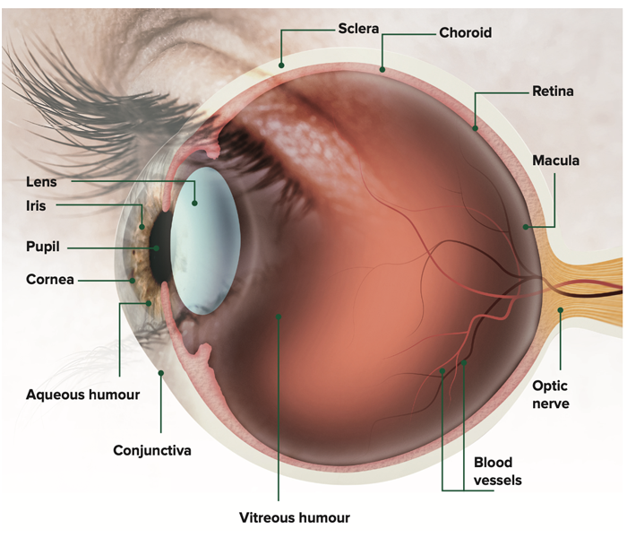
Different parts of the eye structure include:
- Cornea – the clear membrane covering the front of the iris and pupil, which refracts the light entering the eye. It covers one sixth of the eyeball’s surface
- Sclera – the tough, protective white covering of the eyeball which is continuous with the clear cornea. It makes up five sixths of the surface of the eyeball, serving as the attachment for the ocular muscles that move the eye
- Conjunctiva – the mucous membrane covering the front part of the sclera and lining the inside of the eyelids. It protects and lubricates the eye
- Choroid – the spongy middle layer of the eyeball located between the sclera and the retina. It provides oxygen and nourishment to the outer layers of the retina
- Aqueous humour – a watery fluid behind the cornea that circulates in the front part of the eyeball and makes sure the pressure inside stays constant
- Iris – the coloured circle surrounding the pupil which changes the size of the pupil to allow varying amounts of light into the eye
- Pupil – the dark, circular hole in the centre of the iris through which light enters the eye. It tends to get larger in dim light and smaller in bright light
- Lens – a transparent structure behind the iris that changes shape to bend light rays, helping to focus them onto the retina
- Vitreous humour – a colourless, gel-like material filling the inside of the eyeball from the lens to the retina, helping to keep the eye inflated
- Retina – a layer of light-sensitive cells right at the back of the eyeball that work by collecting and sending information from light signals along the optic nerve
- Macula – the small and highly sensitive bullseye at the centre of the retina responsible for detailed central vision
- Optic nerve – takes information from the retina to the brain
- Blood vessels – bring nutrients to the nerve cells.